New science, technology, and dedication improve dermatopathology
Dedication and attention to detail by dermatologists enhance the effectiveness of new developments.

New evidence and approaches are bringing important changes to dermatopathology. These include screening for genetic syndromes using immunohistochemistry, molecular testing, new immunochemistry stains, and new considerations in managing atypical nevi, to list a few changes. What hasn’t changed is the need for clinicians to provide complete biopsy specimens, photos and images, and relevant clinical information.
“We want to be sure we are looking at adequate specimens, the entire lesion,” said Alina G. Bridges, DO, FAAD, director of immunodermatology at the Richfield Laboratory of Dermatopathology. “We need to know that half the lesion isn’t still on the patient and that remnant has melanoma when all we see is a little atypia.”
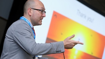
Dr. Bridges will review the latest developments during “Game Changers in Dermatopathology” (U019) Saturday morning. It takes more than new science and technology to change the game of dermatopathology, she said. It also takes dedication and attention to detail by dermatologists.
Patients with a history of dysplastic nevi are at increased risk for developing melanomas, and the melanomas are more likely to develop at a new site, she said. Current guidelines suggest that patients with moderately dysplastic nevi should be followed for later melanomas. But a dermatopathologic finding of moderate dysplasia in a nevus is only as good as the submitted specimen that was assessed.
“If there is any concern that the patient has a highly suspicious pigmented lesion, then the lesion should be sampled as an excisional biopsy with 1-3 mm clear clinical margins rather than a partial biopsy,” Dr. Bridges advised.
Immunohistochemical staining is another important issue. Stains can be expensive, and a barrier to care for patients who are uninsured or have high deductibles. She suggested ordering stains only if there is a need to improve diagnostic capabilities, help with prognosis, and guide therapy.
Some lesions are more suspicious than others. Staining could be helpful for patients with sebaceous adenomas or sebaceous carcinomas and sometimes reticulated acanthomas with sebaceous differentiation. These neoplasms can be associated with Muir-Torre syndrome, a variant of hereditary nonpolyposis colorectal cancer. If immunochemical staining reveals defective DNA mismatch repair proteins, the patient can be referred to medical genetics for further evaluation.
She said dermatopathologists are excited about a new stain called PRAME, preferentially expressed antigen in melanoma, which can help distinguish melanoma from a benign nevus. But like many stains, PRAME is not foolproof. It cannot always identify desmoplastic melanoma, which has the lowest positivity of all melanoma subtypes.
“Sometimes a regular nevus is positive and sometimes an unequivocal melanoma is negative,” Dr. Bridges cautioned. “Many people are bringing it on board, but it is not a replacement for other melanocytic markers.”
There is also a novel immunostain for the Merkel cell polyomavirus. A positive stain is associated with a better prognosis. Loss of BAP1 nuclear immunohistochemistry staining can identify a BAPoma or BAP1 inactivated melanocytic tumor, which can be associated with a BRAFV600E mutation.
Molecular testing is becoming similarly important, Dr. Bridges said. Testing is currently recommended when a diagnosis of melanoma is in question and in cases of indeterminant atypical melanocytic neoplasms.
“Molecular testing adds to the cost, but is almost essential for diagnosis, prognosis, and therapy in these difficult melanocytic neoplasms,” she said. “If, based on testing, for example, an atypical Spitz tumor chromosomal aberrations like melanoma, then we diagnose and manage the lesion as melanoma, although it often has a better prognosis. This is significant because atypical Spitz nevi and tumors can occur in younger patients.”


