The ABCs of dysplastic nevi
These pigmented lesions are not always a pathway to melanoma, so proper planning and counseling is crucial.
Dysplastic nevi are a relatively common occurrence. Despite this, the treatment and management of these atypical moles remain controversial and challenging.
Elizabeth Gates Berry, MD, FAAD, associate professor of dermatology at the Oregon Health & Science University in Portland, said there are three reasons for the difficulty surrounding dysplastic nevi.
“We don’t know the true malignant potential of the histologically dysplastic nevi, they are very challenging to characterize under the microscope, and there is a wide spectrum of approaches to management,” she said. “Due to the inherent uncertainties in the diagnosis, anecdotal evidence and fear of melanoma can play a large role in treatment decisions.”
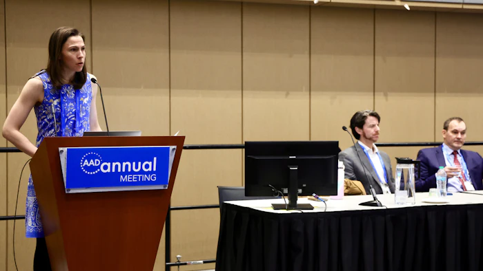
Those decisions were part of the discussion in Monday’s Annual Meeting session, U101 – Dysplastic Nevi for Dummies, directed by Dr. Berry and featuring presentations from Benjamin Stoff, MD, FAAD, and Michael Tetzlaff, MD, PhD.
 Elizabeth Gates Berry, MD, FAAD
“The intra- and inter-observer agreement among pathologists histologically diagnosing dysplastic nevi is poor,” she said. “Because of these uncertainties, many clinicians still elect to re-excise dysplastic nevi of varying grades.”
Dr. Berry said it is critical to remember that dysplastic nevi are not necessarily precursors to melanoma, but rather they are markers of risk. She said recent literature backs this up and points toward changes in the management of dysplastic nevi going forward.
A consensus statement from the Pigmented Lesion Committee, “Addressing the Knowledge Gap in Clinical Recommendations for Management and Complete Excision of Clinically Atypical Nevi/Dysplastic Nevi,” was published in JAMA Dermatology. Dr. Berry said the statement recommends observation for mildly dysplastic nevi with positive or negative margins and for moderately dysplastic nevi with negative margins.
“The group also advised re-excision for severely dysplastic nevi regardless of margin status,” she said. “The subcommittee identified management of moderately dysplastic nevi with positive histologic margins as an area for additional research.”
A study discussed in the session, “Risk of Subsequent Cutaneous Melanoma in Moderately Dysplastic Nevi Excisionally Biopsied but With Positive Histologic Margins,” was also published in JAMA Dermatology and backed up the assertion that dysplastic nevi are markers for risk.
The study analyzed 467 moderately dysplastic nevi with positive margins that had no residual clinical pigment following the biopsy. With a mean follow-up of nearly seven years, no same-site melanomas developed. However, 23% of the patients went on to develop a separate-site melanoma.
“This study highlighted that dysplastic nevi are not obligate precursors to melanoma and supports monitoring over re-excision of moderately dysplastic nevi with positive margins,” Dr. Berry said.
However, there are still some patients who will be candidates for re-excision, she said. These patients were the subject of another study, “Development of a Treatment Decision Aid for Patients With Dysplastic Nevi Who Are Candidates for Re-Excision,” which was published in the Journal of the American Academy of Dermatology.
Dr. Berry, who was one of the researchers on this study, said the team used existing literature to develop a patient decision aid (PDA) to help guide discussions of treatment options with patients. The team is working on another paper that evaluates a quantitative (containing estimates of melanoma development) versus qualitative (descriptions only) version of that PDA.
“Overall, study participants preferred the quantitative version,” Dr. Berry said. “Interestingly, approximately 30% of the participants selected a treatment that differed from expert recommendation, which underscores the importance of shared decision-making.”
 Benjamin Stoff, MD, FAAD
“Techniques like FISH, gene expression profiling, comparative genomic hybridization, and next generation sequencing can be used selectively to aid dermatologists in determining the likelihood that an ambiguous melanocytic lesion represents melanoma,” Dr. Stoff said, adding that guidance from both the National Comprehensive Cancer Network and the American Society of Dermatopathology support the use of molecular techniques in this context.
Dr. Tetzlaff, who is a clinical professor of pathology and dermatology in the dermatopathology and oral pathology unit at the University of California, San Francisco, agreed with Dr. Stoff, adding there are some specific scenarios in which molecular testing can be of value.
“If the differential diagnosis is between a severely atypical dysplastic melanocytic nevus and melanoma, with some features resembling a dysplastic nevus, then I think molecular testing can be informative in select cases,” Dr. Tetzlaff said. “The test deployed often depends on the amount of cellularity present in the biopsy. FISH and TERT promotor sequencing are often most accessible. If the lesion is sufficiently cellular, next generation sequencing, which in our hands provides information regarding copy number alterations and somatic mutations, could be deployed.”
Ultimately, Dr. Berry said that recent consensus recommendations and studies suggest that monitoring alone is safe for mild or moderate dysplastic nevi regardless of margin status — with the caveat that the nevus is biopsied in its entirety without residual clinical pigment.
“There is enough uncertainty surrounding the label of severely dysplastic nevi that most clinicians recommend re-excision for complete removal,” she said. “However, management of severely dysplastic nevi is an area of active research, which may change the clinical recommendations in the coming years.”
Despite what those recommendations may end up being, Dr. Berry said it’s important to remember that every mole is not a “ticking time bomb,” and that the decision to biopsy nevi should be approached carefully.
“Leverage available tools and data to help with the decision to biopsy, including clinical behavior of the nevus, ugly duckling sign, whole body photography, digital dermoscopy, and advanced imaging. And when possible, biopsy the entire nevus,” she said. “If a partial biopsy shows clinical atypia of any type, consider excisional therapy.”
Lastly, Dr. Berry said the patient should be included as much as possible in whatever plan is made.
“Shared decision-making with the patient is key to ensure the patient’s values and preferences factor into the management of the dysplastic nevus,” she said.