Tried, true, and new
AAD offers the opportunity for all dermatologists to learn or cultivate skills in cutaneous surgery.
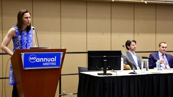
 Elizabeth Billingsley, MD, FAAD
3:30-5:30 p.m. | Monday, March 30
Bluebird 2A
Any dermatologist can benefit from continuing education to strengthen foundational surgical skills in cutaneous procedures. Tomorrow, that prospect comes to fruition in the new Annual Meeting session, F101 – Basics of Cutaneous Surgery.
The session will cover the full arc of dermatologic surgery — from planning and executing excisions to instrumentation, suturing techniques, and postoperative management. The six-person panel will deliver a thorough, practical review of essential cutaneous surgery principles, said session director Elizabeth Billingsley, MD, FAAD, a professor of dermatology at the Penn State University College of Medicine and Penn State Health in Hershey.
 Franki Lambert Smith, MD, FAAD
Session speaker Franki Lambert Smith, MD, FAAD, underscored the session’s central mission: to help all dermatologists feel confident in all aspects of dermatologic surgery, including case selection, reconstruction, and complication management. Dr. Lambert Smith, an associate professor of dermatology at the University of Rochester Medical Center in New York, said speakers will provide individual tips and tricks that can help physicians perform successful surgeries.
First things first
 Norhan Shamloul, MD, FAAD
- The temporal branch of the facial nerve (CN VII) is vulnerable as it crosses the zygomatic arch; injury can result in eyebrow ptosis.
- The marginal mandibular branch (CN VII) is located at the angle of the mandible; injury can cause facial asymmetry and uneven smile.
- The spinal accessory nerve (CN XI) is present in the posterior triangle of the neck; injury can cause chronic shoulder pain and inability to abduct the shoulder.
“These anatomical landmarks underscore the importance of precision and awareness in surgical planning,” Dr. Shamloul said.
Precise cut
Amrit Greene, MD, FAAD, an assistant professor of dermatology at Geisinger Medical Center in Danville, Pennsylvania, will explore tips for excisions. Designing an excision requires consideration of multiple factors, he said, including tumor characteristics that determine appropriate margins, anatomic location, relaxed skin tension lines, and anticipated closure tension.
According to Dr. Greene, the process begins by outlining an elliptical excision encompassing the lesion with appropriate margins, typically using a length-to-width ratio of approximately 3:1 to facilitate linear closure.
“The long axis of the ellipse is oriented parallel to relaxed skin tension lines or within natural anatomic creases, when feasible, to minimize scar visibility. The incision is carried through to the subcutaneous tissue, with depth dictated by tumor characteristics, and perpendicular wound edges are maintained,” he said.
Additionally, Dr. Greene said the extent of undermining is guided by the tension of the resulting defect and patient-specific bleeding risk factors, followed by meticulous hemostasis using electrocautery or similar methods.
“Closure is generally performed in layers, with deep absorbable sutures to reduce tension and superficial sutures or adhesive techniques to achieve precise epidermal approximation and optimal cosmetic outcomes,” he said.
In high-tension areas, Dr. Greene said specialized suture techniques may be employed to further offload epidermal tension, and in select cases, full-thickness skin grafts or local flaps are utilized to preserve function and structural integrity of surrounding tissues.
Big finish
The new session will also include a discussion on flap design and execution, led by Kathryn M. Potter, MD, FAAD, an associate professor of dermatology at Augusta University’s Medical College of Georgia. Dr. Potter will address when flaps are necessary in surgical repair — a common decision point following skin cancer removal, especially in Mohs surgery. As a Mohs surgeon specializing in the head and neck, she frequently employs various flap techniques to close defects created during cancer excision.
 Kathryn M. Potter, MD, FAAD
Dr. Potter noted that her talk is not centered on new devices or breakthrough techniques; instead, it reinforces the tried‑and‑true approaches that remain the backbone of cutaneous reconstruction. Precision, she said, outweighs novelty when it comes to flap design. Understanding tissue tension, ensuring adequate blood supply, and avoiding pitfalls such as “pin cushioning” are core skills she aims to reinforce. Broad undermining, for example, can help reduce distortion and improve healing.
Caution ahead
In terms of dermatologic surgery complications, Kimberly Lai Brady, MD, FAAD, said that although such instances are infrequent and minor, they can be further minimized by recognizing early warning signs and taking preventive steps during the surgical process. Dr. Brady is a Mohs surgeon at Roswell Park Comprehensive Cancer Center in Buffalo, New York.
 Kimberly Lai Brady, MD, FAAD
Dr. Brady further noted that clean, dehisced wounds may be re-sutured or may be left to heal by granulation. Hematoma development often occurs within the first 48 hours after a surgical procedure and appears as tense, painful swellings under a closed wound, which can lead to overlying skin necrosis.
“Patients on anticoagulants have a higher risk of hematoma development and postoperative bleeding. Meticulous hemostasis with electrosurgery during the procedure, limited undermining, and pressure dressings can reduce the risks of postoperative bleeding complications,” she said. “When complications arise, recognition is key and management should be prompt to ensure optimal healing and outcomes.”