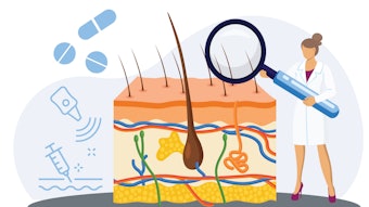
Enhancing the naked eye with medical imaging
Medical imaging advances dermatology.
Photographic images have long been a useful record of dermatologic changes. Advancing technology — combined with growing awareness of the value of clinical imaging — is transforming a useful record into a must-have, must-use clinical tool.
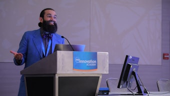
“Just as a radiologist can diagnose breast cancer from a mammographic image seen through a monitor, dermatologists, who are by far the most visual of medical specialists, need to learn and do likewise,” said Paola Pasquali, MD, IFAAD, coordinator of dermatology at Pius Hospital in Valls, Spain.
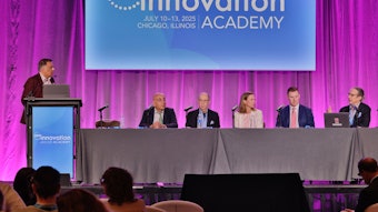
Dr. Pasquali moderated the session F067 – How Far Can We Get With Medical Imaging and explored how far imaging technology has already taken dermatology and how the practice is likely to evolve. For far too long, dermatologists have relied on eyesight and paid too little attention to imaging modalities developed by other specialties.
Beyond the clinical eye
“A radiologist or an endoscopist would be thrilled if you offered a new gadget to their armamentarium that would allow them to improve their diagnostic capacities,” Dr. Pasquali noted. “Dermatologists have relied on their eyes. Just imagine the change in the treatment of keratinocyte tumors if we know beforehand the depth and shape.”
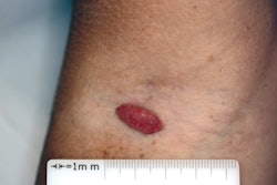
Dermatologists have been using photographs to document, diagnose, monitor, and follow up on treatment outcomes since glass plates were the newest technology in the 19th century. Photographs have been familiar teaching tools for nearly two centuries, a role that continues to grow as new imaging modalities come into clinical practice.
Advances in dermoscopy
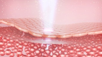
Dermoscopy is the second most common imaging modality in dermatology, Dr. Pasquali continued. From the early use of surface microscopy in the 1920s, dermoscopy now allows dermatologists to magnify and visualize subepidermal structures. High-frequency ultrasound, reflectance confocal microscopy, and optical coherence tomography (OCT) are bringing deeper and more detailed views into practice.
“The naked eye is simply not sufficient,” Dr. Pasquali said. “We need to go beyond the visible and obtain as much information as possible to make more precise diagnoses and make the most appropriate decisions for each condition.”
Adopting new technologies
All of these novel imaging approaches meet real medical needs, she said. Some clinicians use confocal microscopy as a noninvasive biopsy, an in vivo assessment of cellular morphology. Ultrasound and OCT provide detailed information about the volume, shape, and depth of invasion of tumor diseases. Those same approaches can be used to more accurately diagnose and monitor inflammatory reactions to aesthetic fillers.
Cost is always a consideration in adopting new technologies, Dr. Pasquali added. New imaging modalities are no exception. One approach is to incorporate multiple imaging techniques into one piece of equipment: photography and ultrasound, confocal plus photography plus dermoscopy.
“The synergistic effects of combining two or more techniques enhances their diagnostic impact and practicability,” she said. “Excellent cameras are already part of mobile phones, and attachments for mobiles make it possible to use and take images from any brand of dermoscope. There is already a prototype for a mobile confocal microscope — it is just a matter of time.”
Visit AAD DermWorld Meeting News Central for more articles.